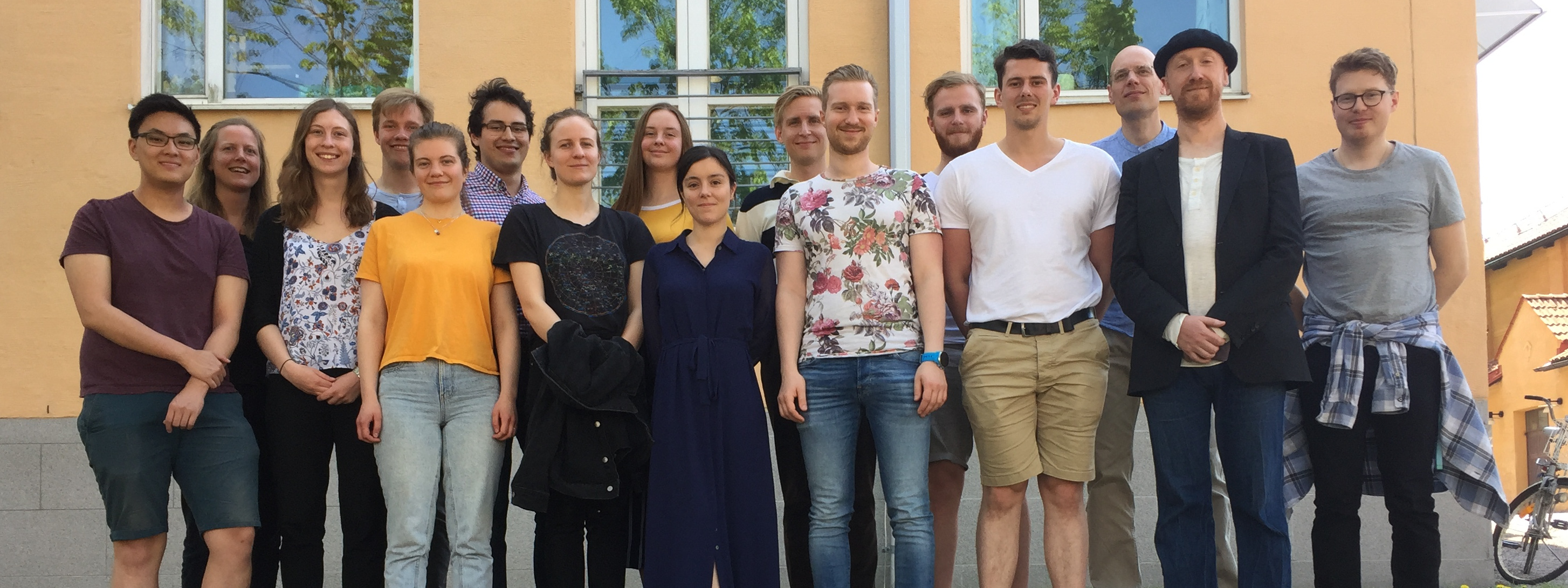
Today there is a new Ph.D. defense in ISB group: that of Markus Karlsson. Markus has been working with usage of magnetic resonance imaging techniques to characterize the liver, and the formal title of the Ph.D. thesis is: “Non-Invasive Characterization of Liver Disease – by multimodal quantitative magnetic resonance”. Various techniques have been tested, MRS-PDFF (to measure liver fat), T1 relaxation (to estimate fibrosis), R2* (to estimate iron in the liver), etc. In three of the papers (Paper 1,2 and 4), normal MRI analysis has been done in different patient cohorts, and in two of the papers (Paper 3 and 5), mathematical modelling has been done in collaboration with ISB group. More specifically:
Paper 1 established a relationship between R2* and MRS-PDFF, and saw that liver fat distorts the R2* signal with about one R2* unit per MRS-PDFF unit. This could be used to device a simpe correction method when using R2* to estimate liver iron levels.
Paper 2 looked at the relationship between T1 and liver fibrosis in a cohort of approximately 100 patients with various degrees of diffuse liver disease, ranging from no fibrosis to cirrhosis. In the literature, different degrees of connection between T1 and liver fibrosis have been reported, and this paper unfortunately supports the conclusion that there is low correlation.
Paper 3 was the main paper deviced by us:
Model-inferred mechanisms of liver function from magnetic resonance imaging data: Validation and variation across a clinically relevant cohort. Forsgren MF, Karlsson M, Dahlqvist Leinhard O, Dahlström N, Norén B, Romu T, Ignatova S, Ekstedt M, Kechagias S, Lundberg P, Cedersund G. PLoS Comput Biol. 2019 Jun 25;15(6):e1007157.
In this paper, the data consists of DCI-MRI data, i.e. dynamic MRI images which have been enhanced by the contrast agent gadoxetate, which has been injected into the arm at the beginning of the time-series. By combining this data with a previously developed compartment transport model, we could estimate reliable uptake rates into the liver for all patient groups. Furthermore, in this paper we also showed that these estimated uptake rates, only accesible using the model, serve as new useful biomarkers for estimating liver function and fibrosis. In this paper, we also demonstrated how nonlinear mixed-effects modelling could be used to simplify the currently used protocol, to save money and time in the clinic.
Paper 4 looked at the relationship between magnetic resonance cholangiopancreatography (MRCP) and liver function estimated using DCI-MRI.
Paper 5 again looks at modelling of DCI-MRI data, to characterize the transport rates in and out of hepatocytes. In this paper, we compare uptake rates in humans and rats, and in rats with different exposures to a drug which impacts liver function. This allows us to establish a translational framework, that can predict the likely effect of a drug in humans, based on these available data and new data for the effect of the drug in rats. This is useful for drug development, when one wants to estimate the likely effect of a drug in humans based on available pre-clinical and clinical evidence.
Opponent for the defense is Steven Sourbron, who has worked with similar uptake and perfusion modelling in many of the central organs for many years, and who is one of the leading authorities in the field. In the examination committee sits Sven Månsson (Malmö), Lennart Blomqvist (Stockholm) and Zoltan Szabo (Linköping), who have complementary radiological and clinical competence.

